
Key takeaways
- Anaerobic culture finds germs that die in air and that normal cultures miss.
- The sample is sealed away from oxygen from the very first second; deep aspirates beat surface swabs.
- Preliminary reports take 48 to 72 hours; full susceptibility takes three to five days.
- The S/I/R table guides your doctor's antibiotic choice; it is not a prescription.
- Tell your doctor about any antibiotics already taken before the sample is drawn.
- No growth is usually good news; a late or antibiotic-wiped sample can also come back empty.
Book tests from this article
Sample collected at home - free
- Anaerobic Culture and Susceptibility (Manual method)
₹1,849₹2,24918% off
- Anaerobic Culture and Susceptibility(Vitek 2 Compact)
₹2,099₹2,79925% off
- Blood Culture And Sensitivity (Anerobic)(Vitek 2 Compact)
₹1,699₹2,24924% off
Free collection 24 hr reports
The one-paragraph version
An anaerobic culture and susceptibility (manual method) test is the lab's way of catching bacteria that die when they touch air. Your doctor orders it when a wound, abscess, or deep infection is not healing on routine antibiotics and the regular culture came back with nothing useful. The sample (usually pus or fluid drawn from deep inside the infected area) is sealed away from oxygen from the very first second, grown in an airtight jar for 48 to 72 hours and sometimes up to a week, then tested against a panel of antibiotics to produce the S/I/R grid your doctor uses to pick treatment. It is slow on purpose. Rushing it gives a wrong answer, and a wrong answer here wastes more of your time than the wait ever could.

Why this test exists when the routine culture came back empty
Most culture requests you will ever see are aerobic. The swab goes onto a plate, the plate sits on a lab bench in ordinary room air, and whatever grows, grows. That works fine for the bacteria that like air, which is most of the troublemakers you hear about. But a whole family of germs, the anaerobes, cannot tolerate oxygen at all. Some die within minutes of exposure. Others go dormant and never wake up again on the plate.
So here is the frustrating pattern patients describe to us at the counter in Malad. The wound looks infected: red edges, swelling, pus. The doctor sends a routine culture. The report comes back "no growth" or grows only skin bacteria that do not explain the picture. Antibiotics are prescribed anyway, the full course is finished, the dressing gets changed, and the wound looks exactly the same. Nobody made a mistake. The test just asked the wrong question. A bench-top culture in room air can never find a germ that oxygen kills. That is the entire reason the anaerobic request exists.
It is worth knowing that this is not a rare edge case. Anaerobic bacteria are everywhere in the human body. Your gut carries more of them than any other kind of bacterium. They live on your gums, in your throat, and throughout the lower intestine, mostly as harmless neighbours. They only turn dangerous when they escape into places they do not belong: deep inside a wound, behind a blocked duct, inside an abscess wall, or into the bloodstream after surgery or trauma. When that happens, the infection is usually a team effort. Anaerobic infections are very often polymicrobial, meaning several germs at once, both anaerobic and aerobic, which is one more reason a single routine culture can miss the real story.
What "anaerobic" actually means
The word gets thrown around as if everyone learned it in school, so let us pin it down. An obligate anaerobe is a bacterium that cannot grow in the presence of oxygen. Some are poisoned by it outright. Their enzymes cannot handle the reactive oxygen species that air exposure creates, so the cells die. A smaller group, the aerotolerant anaerobes, can survive a whiff of air but still only grow properly without it. For the lab, the practical difference barely matters. Either way, the sample must be protected from air from the moment it leaves your body, or there is nothing left to grow.
The names you are most likely to see on a report belong to a short list. Bacteroides, especially Bacteroides fragilis, is the single most common cause of anaerobic infection in humans and a familiar face in abdominal infections and abscesses. Fusobacterium turns up in head and neck infections. Prevotella and Porphyromonas cluster around dental and gum infections. Clostridium species are the ones behind gas gangrene and some of the nastiest wound infections. Peptostreptococcus and Actinomyces round out the usual suspects. You do not need to memorise this list. What matters is the pattern: each of them lives naturally in some dark, airless pocket of the body, and each causes trouble only when it escapes that pocket.
Clinically, anaerobic infections have a couple of telltale signs that doctors learn to recognise. One is the smell. Pus from an anaerobic infection often carries a distinctly foul, putrid odour that experienced clinicians notice immediately. The other is the abscess itself: a walled-off pocket of pus is exactly the kind of low-oxygen environment where anaerobes thrive, which is why drainage of an abscess matters as much as the antibiotic that follows. Antibiotics alone often cannot finish the job inside a sealed pocket of pus, because drugs penetrate abscess walls poorly and the acidic, oxygen-free interior inactivates some of them. That is why surgeons say source control, meaning draining the pus and cleaning out dead tissue, is half the treatment. The culture tells the doctor which drug to use; the drainage gives the drug a fighting chance.
One nuance that saves a lot of confusion later: finding an anaerobe on a report does not always mean it caused your infection. These germs are normal residents of the gut, mouth, and skin in small numbers, so a swab dragged across the surface of a chronic ulcer can grow Bacteroides that were simply living there harmlessly. Microbiologists and doctors read the result against the source. The same organism grown from pus aspirated out of a sealed abdominal abscess means infection and needs treatment. Grown from a surface swab of a diabetic foot ulcer, it may mean nothing at all. The specimen source is not a footnote on the form. It is half the diagnosis.
The vial: the first sixty seconds after collection
This is not a blood test, and there is no needle in your arm. The sample is the infection itself: pus, wound fluid, or a piece of infected tissue. How it is taken depends on where the infection sits, and the method matters more here than in almost any other culture.
The gold standard is aspiration. For an abscess, the doctor numbs the skin with a small local anaesthetic injection, makes a tiny nick or passes a needle directly into the pocket, and draws the pus into a syringe. Patients always ask about the pain, so here is the honest version: the numbing shot stings for a few seconds, sharp like a bee sting, and after that you feel pressure and tugging but not real pain. The whole thing is over in a minute or two. For a deep wound, the doctor or nurse takes a swab or a small piece of tissue from the base and walls of the wound, not from the surface. That distinction is everything. Anaerobes live in the depths where oxygen cannot reach. A swab wiped across the top of a wound collects skin bacteria and air, and it comes back with the wrong answer. Deep sampling is the difference between a useful report and a wasted week.
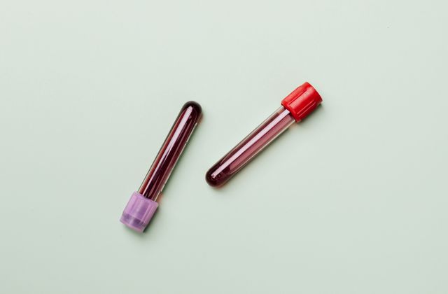
Then comes the part you never see but the lab obsesses over: the transfer. The syringe or swab goes straight into a sealed anaerobic transport vial, a small container designed to keep oxygen out. Air is pushed out of the syringe first, the cap goes on, and from that second onward the clock we will talk about next is already running.
At our lab, we tell every patient the same thing: if the sample spends too long outside the body, we would rather ask for a repeat than report a misleading "no growth." A dead germ tells us nothing, and a wrong "no growth" can send your doctor down the wrong path for weeks.
Tissue biopsies, when the doctor takes a small piece of infected tissue instead of fluid, are excellent samples too. They carry more bacteria and less surface contamination than any swab. Sterile body fluids, fluid from a joint, pleural fluid from around the lungs, and aspirates from deep neck or pelvic collections can all be cultured anaerobically as well. What the lab does not want is a dry swab from a superficial scrape, or a sample scooped from the skin surface around the wound. Those are the specimens most likely to grow the wrong germs and miss the right ones.
The clock starts ticking the moment the sample leaves your body
Anaerobes do not wait politely. From the instant the sample is exposed, oxygen starts killing the very organisms the test is trying to grow. That is why transport rules for this test are stricter than for nearly anything else in the lab, and why "the sample reached late" is one of the most common reasons for a repeat request.
The rule of thumb depends on the container. A swab in a basic transport tube is fragile cargo: the lab wants it within a couple of hours at room temperature, and refrigeration does not help. Cold slows some bacteria down, but it damages many anaerobes and encourages the growth of contaminants that then overgrow the plate. So the instruction is simple: room temperature, fast, no fridge. A sample drawn into a proper anaerobic transport vial, where oxygen has been excluded from the start, is sturdier. Sealed correctly, it can hold for up to about 72 hours at ambient temperature and still be worth culturing. That is the window reference laboratories quote, and it is the reason the vial itself matters as much as what is inside it.
This is also why our riders move the way they do. When we collect a pus or wound sample from a home in Malad, Kandivali, Borivali, or Goregaon, the technician carries the sealed anaerobic vial back to the lab on the same run, not in a batch at the end of the day. The lab itself runs 24x7, which matters more for this test than for most: a sample taken at 9 in the evening does not sit in a corner until morning. It goes onto plates the same night, because every hour in transit is an hour of oxygen exposure, and the germs we want are dying while we talk about it.
There is one more timing rule, and patients push back on it the most. Ideally, the sample is taken before antibiotics start, or at least before the next dose. Antibiotics in your bloodstream also reach the wound, and they can sterilise the sample: kill or suppress the bacteria so thoroughly that nothing grows, even though the infection is real. Sometimes that cannot be avoided. If you are already on antibiotics and the doctor still orders the culture, the timing is adjusted around your doses and the lab reads the result with that in mind. But if nobody told the doctor you were already taking something, the lab is flying blind. Tell them. It is the single most useful sentence you can say at the time of collection, after your name.
A week inside the anaerobic jar
Once your sample reaches the lab, it enters a small world with no air in it. The microbiologist smears the material onto agar plates and seals them inside an anaerobic jar: a heavy, airtight container where a chemical sachet reacts to scrub out every trace of oxygen and replace it with a gas mix the anaerobes prefer. Some laboratories use anaerobic chambers instead, which are glove-box cabinets filled with oxygen-free gas where the microbiologist works through sealed sleeves. The principle is identical. From this point on, nothing about the sample touches ordinary air until the reading is done.
Then the waiting starts, and it is longer than most patients expect. The sealed plates go into an incubator at body temperature, around 35 to 37 degrees, and stay there for at least 48 to 72 hours before the first real reading. Slow growers may need up to seven days. This is not the lab being lazy. Anaerobes simply divide more slowly than their aerobic cousins, and colonies that are invisible on day two can be obvious on day four. A preliminary report often goes out at the 48 to 72 hour mark, saying what has grown so far, and the final report follows when the plates have had their full time.
What does the microbiologist actually do when the jar is opened? The honest answer is part science, part craft. Each colony is studied by eye: its size, shape, colour, edge, and surface texture. Some are even identified partly by smell, because certain anaerobes produce unmistakable odours as they metabolise. A Gram stain comes early, colouring the bacteria purple or pink and revealing rods or spheres under the microscope, which narrows the field enormously. Then small biochemical tests confirm the identification: does this organism ferment this sugar, does it produce that enzyme. Organisms like Bacteroides and Fusobacterium each have their own look on the plate, and an experienced microbiologist can often make a good guess before any instrument confirms it. Automated systems exist, and you may see the Vitek 2 Compact version of this test on some request slips, but the manual method is the same biology with more hands-on reading.
There is a quiet discipline to this work that patients never see. Every time the jar is opened to check the plates, oxygen rushes in, so readings are quick and the jar is resealed fast. Plates that dry out or get contaminated during a check are useless. The senior microbiologists who do this well have usually been reading anaerobic plates for years, and their eyes are calibrated in a way no manual can fully teach. When your report takes five days, some of that time is literally a person looking at your colonies, twice a day, waiting for them to declare themselves.
The susceptibility step: what the S/I/R grid really is
Growing the germ is only half the test. The other half answers the question your doctor actually needs: which antibiotic will kill it. That is the susceptibility part, and the grid of S, I, and R letters on your report is its entire output.
Here is how it works on the bench. Once the lab has a pure growth of your organism, it prepares a fresh plate spread evenly with that bacterium and exposes it to a panel of antibiotics. In the classic manual method, this looks like small paper discs, each soaked in a fixed amount of one antibiotic, placed on the agar surface. The plate goes back into the oxygen-free environment. Where an antibiotic works, it diffuses outward and leaves a clear zone with no bacterial growth around the disc. Where it fails, the bacteria grow right up to the disc's edge. The microbiologist measures those clear zones in millimetres and compares them against published breakpoints, which are cutoff values set by standards bodies such as the CLSI. A zone at or above the breakpoint reads Sensitive. Below a lower cutoff reads Resistant. In between sits Intermediate. (Reference laboratories often confirm with dilution methods, where the antibiotic concentration is varied in tubes or wells to find the minimum inhibitory concentration, but the disc reading you picture is the workhorse of routine labs, and it is an honest simplification of what happens.)
Let us walk through a miniature example, because this is the part of the report that confuses everyone. Imagine your report lists three antibiotics against the grown organism. The first shows S. That means the antibiotic stopped the organism's growth at concentrations the drug normally reaches in the body, so it is a reasonable choice. The second shows I, intermediate. That does not mean "half works." It means the drug might work only at higher doses or in body sites where the drug concentrates well, like the urinary tract, and your doctor will usually prefer an S option if one exists. The third shows R. That one failed outright in the lab, and prescribing it would be guessing against the evidence. Your doctor reads this grid alongside everything else they know about you: how sick you are, if tablets will do or an IV is needed, what you have taken before, and whether the infection needs drainage as well as drugs.
Two things about this grid deserve emphasis. First, it is guidance for the prescriber, not a shopping list. An S does not mean "buy this yourself." Antibiotic choice involves your kidney function, allergies, other medicines, and the infection's location, none of which the lab can see. Self-medicating from a susceptibility report is one of the fastest ways to breed resistance and still stay infected. Second, the grid only tests the antibiotics on the panel. If your doctor wants to try something not listed, that drug was not tested against your germ, and the report says nothing about it either way.
Why does the susceptibility step add days to the timeline? Because it is a second full round of growing. The organism must first be identified, then regrown in pure culture, then exposed to the antibiotics, then incubated again under anaerobic conditions for another 48 to 72 hours before the zones can be read. That is why the final susceptibility report lands at three to five days even when the preliminary culture report arrived earlier. Each stage depends on the one before it, and bacteria do not negotiate deadlines.
Reading your report line by line
Reports vary by laboratory, but the anatomy is nearly always the same, and it helps to know which line is which before you open the PDF with shaky hands.
The first section is the specimen line. It should say what was sent and where it came from: "pus, aspirated from abscess, right thigh," for example. Check that this matches you. A wrong specimen source on the form can change how the whole report is interpreted, as we discussed: the same organism means different things in different samples.
Next comes the Gram stain result, if your lab reports it. This is the early microscopic look, usually available well before the culture finishes. It will say something like "many pus cells seen, gram-negative rods seen" or "no organisms seen." Pus cells are your own white blood cells, and their presence confirms there is real inflammation at the site. "No organisms seen" on the Gram stain does not contradict a later positive culture; the stain needs a fairly high number of bacteria to spot them, while the culture can grow from far fewer.
Then the culture result itself: the name of what grew, often in italics, with a comment on the amount. "Heavy growth of Bacteroides fragilis" is a clear, significant result. "Scanty growth" or "light growth" means fewer colonies, which the microbiologist weighs against the specimen source before calling it significant. "Mixed anaerobic flora" means several anaerobes grew together without one clear dominant organism. In a deep, properly collected sample, that usually reflects a genuinely polymicrobial infection, which is common with anaerobes. In a surface swab, it more likely reflects contamination with normal flora, which is why the collection method keeps coming up.
Finally, the susceptibility grid: rows of antibiotics, each marked S, I, or R, sometimes with the measured zone sizes or minimum inhibitory concentrations alongside. Read it as your doctor's menu of evidence-backed options, not as instructions. And remember the laboratory's quiet disclaimer that runs under every culture report: ranges, breakpoints, and methods vary between laboratories, so a result from one lab is interpreted against that lab's own standards.
If your doctor ordered cultures on blood rather than wound fluid, the companion test to read about is the blood culture and sensitivity with anaerobic bottle, which hunts the same kind of germs in the bloodstream and has its own timing and interpretation. And if the slow grower your doctor is chasing lives in body fluid rather than pus, tuberculosis for instance, the AFB DNA TB PCR test on body fluid takes a completely different route, looking for the germ's genetic material instead of growing it.
The three reasons a report says "no growth"
"No growth" is the result patients fear most, because it feels like a dead end. In practice it means one of three things, and only your doctor can tell which one applies to you.
The first and happiest reason is that there genuinely was nothing significant to grow. The inflammation was not bacterial, or the bacteria present were too few to matter, or your immune system had already cleared them. A "no growth" after a properly collected, promptly transported sample from a truly infected site is genuinely reassuring, and doctors do act on it: it steers them away from more antibiotics and toward other explanations.
The second reason is that the sample died in transit. This is the one the lab worries about most, because it looks identical to the first reason on paper. If the swab sat too long, if it was refrigerated, if the vial was not properly sealed, or if the sample was collected from the surface instead of the depths, the anaerobes may have perished before the plates were even poured. A "no growth" that does not match the clinical picture, a wound that is clearly infected but a report that says nothing grew, is exactly when doctors ask for a repeat with better collection. This is not the lab covering a mistake. It is the lab refusing to let a dead sample make medical decisions.
The third reason is antibiotics. If you were already on antibiotics when the sample was taken, the drug may have suppressed the bacteria below the level the culture can detect, even though the infection is still smouldering in the tissue. This is the "wiped clean" sample: real infection, sterile plate. Doctors handle this by timing the next sample before a dose, or by reading the negative result with appropriate scepticism and treating on clinical grounds.
Notice what all three have in common: "no growth" is never the end of the story by itself. It is a piece of evidence that your doctor weighs against what they see with their own eyes. A report never overrules the patient sitting in front of the doctor.
Questions patients ask at the counter
How is an anaerobic culture different from a regular culture? A regular culture grows on an open lab bench in normal air, which kills anaerobic germs within minutes. An anaerobic culture seals everything away from oxygen from the moment the sample is taken: sealed transport vial, anaerobic jar or chamber, oxygen-free incubation for days. Same idea, opposite atmosphere.
Why did my doctor order this when my earlier antibiotics failed? Because treatment without a culture is an educated guess, and the guess assumed the usual aerobic suspects. If anaerobes are driving the infection, the routine culture often returns "no growth," the standard antibiotics miss them, and the wound stalls. The anaerobic culture and its susceptibility grid replace the guess with evidence.
Does the sample collection hurt? It is not a blood draw. For an abscess, the area is numbed first, so you feel the sharp sting of the numbing injection for a few seconds and then pressure and tugging while the pus is drawn. For a deep wound, the swab or tissue sample comes from the base of the wound, which can be uncomfortable for a moment but is over quickly. If a sample ever feels truly painful, say so; the doctor can add more local anaesthetic.
How long does the report take? Longer than any blood test you have had. Expect a preliminary culture report at 48 to 72 hours and the final susceptibility report at three to five days, sometimes longer for slow growers. The lab cannot speed up bacterial division. A lab that promises this result tomorrow is a lab you should worry about.
What does "no growth" mean on my report? Usually good news, the first of the three reasons above, but with the two caveats from that section: if the sample reached the lab too late, or antibiotics taken beforehand wiped the germs out, "no growth" is a dead sample talking, not your body. If your wound is clearly still infected and the report says no growth, your doctor will likely repeat the test rather than accept the result at face value.
What does "mixed anaerobic flora" mean? Should I worry? It means several anaerobic organisms grew together. In a deep aspirate from an abscess or a properly collected tissue sample, that is typical of these infections, which are often polymicrobial, and your doctor treats the pattern. In a surface swab, it more likely means the sample picked up normal flora on its way in. The meaning depends entirely on how the sample was collected, which is why the collection method is printed on the form.
My report lists S, I, and R. Can I choose the antibiotic myself? No. The grid is evidence for your doctor, not a prescription pad. Choosing between the S options involves your allergies, kidney function, other medicines, pills versus an IV, and the infection's location. Picking one yourself risks an ineffective drug, side effects, and resistance that makes the next infection harder to treat.
Why did the lab ask for a repeat sample? Almost always one of three reasons: the sample arrived too late or was transported badly, the swab came from the surface instead of the depths, or you had taken antibiotics that likely sterilised the sample. A repeat is the lab protecting you from a misleading result, not a sign that something went wrong with you.
Can I get this test done at home? Yes, with one caveat that actually matters. Pus and deep wound swabs cannot travel in an ordinary bag; they need sealed anaerobic transport vials and a fast trip to the lab. Our technician brings the vials to your door, collects or receives the sample in the sealed container, and carries it straight back to the lab on the same run. That chain is the whole point of the test, so we do not break it. Bookings are on 84250 40353, and the same routes cover Malad, Kandivali, Borivali, and Goregaon every day.
Why we ask which antibiotics you have already taken
Every anaerobic culture form has a small box for "antibiotics taken," and patients routinely leave it blank because it seems irrelevant. It is the most clinically important box on the form.
Antibiotics do not politely wait outside the sample. Whatever is circulating in your blood is also present in the wound fluid the doctor draws into the syringe, and it keeps working inside the transport vial all the way to the lab. A bacterium that would have grown happily on the plate arrives pre-poisoned and never forms a colony. The report says "no growth," the doctor concludes the infection is not bacterial or has resolved, and the real infection continues underneath. This happens often enough that microbiologists have a mental adjustment for it: a negative culture from a patient on antibiotics is read with far more scepticism than the same result from a patient on nothing.
So the guidance is straightforward. Tell the doctor about every antibiotic you have taken in the past week or two, including the ones you stopped because "they were not working" and the ones a chemist sold you without a prescription. Ideally the sample is drawn before antibiotics begin. When that is impossible, which is often, the doctor times the collection for just before your next scheduled dose, when the drug level in your tissues is at its lowest. Either way, the information lets the lab interpret the result honestly instead of guessing.
This also answers a question we hear a lot: "Can I just stop the antibiotic for a day so the test works?" Do not do this on your own. Stopping antibiotics abruptly can let the infection surge, and the decision to pause or adjust a dose belongs to your doctor, who balances the test's accuracy against your safety. Mention it, let them decide.
What this test cannot do
No test is universal, and this one has honest limits worth knowing before you pin all your hopes on it.
First, it is slow, and some clinical situations cannot wait. A rapidly spreading soft-tissue infection, suspected gas gangrene, or a patient going septic will be treated on clinical judgment long before any culture result exists. The culture then serves as confirmation and course-correction, not as the initial decision. That is normal medicine, not a failure of the test.
Second, not every anaerobe grows in the lab, even under perfect conditions. Some are so fastidious that no routine medium supports them. Others are damaged beyond recovery by the time the sample arrives, no matter how good the transport. A negative culture therefore never proves the absence of anaerobic infection on its own. It is one witness, not the whole trial.
Third, the test identifies bacteria, not the whole story. It will not tell you why the wound got infected, if high blood sugar is slowing the healing (a common issue in diabetic foot infections, where your doctor may pair the culture with a fever profile or blood sugar workup to see the systemic picture), or whether the abscess needs surgical drainage rather than more drugs. The culture answers "which germ and which drug." Everything else still needs a doctor's eyes and hands.
Fourth, methods differ between laboratories. A manual culture read by an experienced microbiologist, an automated system like the Vitek 2 Compact, and a molecular test that hunts bacterial DNA each have different strengths. The manual method this article describes gives you the organism plus its actual, measured drug responses, which is why doctors trust it for treatment decisions. Automated systems are faster at identification. DNA-based tests are faster still but cannot tell you which antibiotic will work, because they detect the germ's presence, not its drug susceptibilities. Your doctor picks the method that fits the question being asked.
Booking it from home
This is the test where logistics genuinely affect the result, so here is how the chain works. Our technician arrives with sealed anaerobic transport vials; the sample goes from the doctor's syringe or swab straight into the vial, the vial is sealed, and it rides straight back to the lab on the same run. Nothing sits in a bag on your dining table waiting for a convenient courier slot. Across Malad, Kandivali, Borivali, and Goregaon, that is the standard route, not a special arrangement.
The lab runs 24x7, so a sample collected in the evening goes onto plates the same night instead of waiting for morning. You will find this test on our tests page, where the price sits next to the booking button, and you can book the collection on 84250 40353. If your doctor's slip mentions the automated version instead, that is the same test run on the Vitek 2 Compact system, and it is listed there too.
The whole test comes down to this: seal the sample from air in the first seconds, sample the depths not the surface, and let the bacteria set the schedule. The S/I/R grid is your doctor's evidence, not a prescription. And if "no growth" arrives while the wound still looks infected, a repeat with better collection is the honest next step.
References
- StatPearls / NCBI Bookshelf, "Bacteroides Fragilis" clinical review (anaerobic infection presentation, specimen collection, and management principles)
- Mayo Clinic Laboratories, "Bacterial Culture, Anaerobic (ANAE)" test definition (acceptable specimens, collection method, transport stability)
- "Identification and Antimicrobial Susceptibility Testing of Anaerobic Bacteria," peer-reviewed review via PubMed Central (CLSI M11 standard, incubation and reading timelines)
- MedlinePlus (NIH), Gram stain and wound culture laboratory test pages (sample collection basics)
Book these tests at home
Live prices · free home sample collection in Malad, Kandivali, Borivali & Goregaon
Anaerobic Culture and Susceptibility (Manual method)
- Report in 120 hrs
- At our Malad West lab
Anaerobic Culture and Susceptibility(Vitek 2 Compact)
- Report in 120 hrs
- At our Malad West lab
Blood Culture And Sensitivity (Anerobic)(Vitek 2 Compact)
- Report in 120 hrs
- At our Malad West lab
Fever Profile
- Report in 48 hrs
- Free home collection
More tests related to this topic
Other Culture & Sensitivity tests you can book with free home sample collection.
AFB - Xpert panel by Combo(MTB /RIF Detection & AFB Culture) - Extrapulmonary Samples
- Report in 120 hrs
- At our Malad West lab
AFB DRUG SUSCEPTIBILITY PANEL 1ST AND 2ND LINE PANEL 10 DRUGS
- Report in 48 hrs
- Free home collection
AFB Rapid culture by MGIT - BAL / Other Respiratory samples
- Report in 120 hrs
- At our Malad West lab
AFB Rapid culture by MGIT - Body Fluid
- Report in 120 hrs
- At our Malad West lab
AFB Rapid culture by MGIT - Pus, abscess and aspirates
- Report in 120 hrs
- At our Malad West lab
AFB Rapid Culture by MGIT - Sputum
- Report in 120 hrs
- At our Malad West lab
AFB Rapid culture by MGIT - Tissue/Biopsy
- Report in 120 hrs
- At our Malad West lab
AFB Rapid Culture by MGIT - Urine
- Report in 120 hrs
- At our Malad West lab
Anaerobic Culture and Susceptibility(Vitek 2 Compact)
- Report in 120 hrs
- At our Malad West lab
Blood C/S - BacT/Alert (with bottle)
- Report in 120 hrs
- At our Malad West lab
Blood C/S - BacT/Alert (without bottle)
- Report in 120 hrs
- At our Malad West lab
Culture & Identification - Anaerobic bacteria, Pus
- Report in 120 hrs
- At our Malad West lab
Get more from the same visit
Health packages that already include these tests, and tests people usually add.
Frequently asked questions
How is an anaerobic culture different from a regular culture?
A regular culture grows on an open lab bench in normal air, which kills anaerobic germs within minutes. An anaerobic culture seals everything away from oxygen from the moment the sample is taken: sealed transport vial, anaerobic jar or chamber, and oxygen-free incubation for days. Same idea, opposite atmosphere.
Why did my doctor order this when my earlier antibiotics failed?
Because treatment without a culture is an educated guess, and the guess assumed the usual aerobic suspects. If anaerobes are driving the infection, the routine culture often returns no growth, the standard antibiotics miss them, and the wound stalls. The anaerobic culture and its susceptibility grid replace the guess with evidence.
Does the sample collection hurt?
It is not a blood draw. For an abscess, the area is numbed first, so you feel the sharp sting of the numbing injection for a few seconds and then pressure and tugging while the pus is drawn. For a deep wound, the swab or tissue sample comes from the base of the wound, which can be uncomfortable for a moment but is over quickly.
How long does the report take?
Longer than any blood test you have had. Expect a preliminary culture report at 48 to 72 hours and the final susceptibility report at three to five days, sometimes longer for slow growers. The lab cannot speed up bacterial division.
What does 'no growth' mean on my report?
Usually good news: nothing significant grew under anaerobic conditions. But it can also mean the sample reached the lab too late or antibiotics taken beforehand wiped the germs out. If your wound is clearly still infected and the report says no growth, your doctor will likely repeat the test rather than accept the result at face value.
What does 'mixed anaerobic flora' mean? Should I worry?
It means several anaerobic organisms grew together. In a deep aspirate from an abscess or a properly collected tissue sample, that is typical of these often-polymicrobial infections, and your doctor treats the pattern. In a surface swab, it more likely means the sample picked up normal flora on its way in. The meaning depends on how the sample was collected.
My report lists S, I, and R. Can I choose the antibiotic myself?
No. The grid is evidence for your doctor, not a prescription pad. Choosing between the S options involves your allergies, kidney function, other medicines, whether you need pills or an IV, and the infection's location. Picking one yourself risks an ineffective drug, side effects, and resistance that makes the next infection harder to treat.
Why did the lab ask for a repeat sample?
Almost always one of three reasons: the sample arrived too late or was transported badly, the swab came from the surface instead of the depths, or you had taken antibiotics that likely sterilised the sample. A repeat is the lab protecting you from a misleading result.
Can I get this test done at home?
Yes, with one caveat that matters. Pus and deep wound swabs need sealed anaerobic transport vials and a fast trip to the lab. Our technician brings the vials to your door, collects the sample in the sealed container, and carries it straight back to the lab on the same run across Malad, Kandivali, Borivali, and Goregaon. Book on 84250 40353.
HWP Diagnostic Editorial Team
Health guides written by the team at HWP Diagnostic, Mumbai, to help you understand your tests before you book. This article is for general education and is not a substitute for medical advice - reference ranges vary between labs, so always discuss your results with your doctor. Read our editorial policy. Photos: Pexels.